

Ultrasound of the pelvic organs
Ultrasound allows you to visualize and assess the condition of the internal organs of the pelvis in women and men. In women, the study includes examination of the uterus, ovaries and fallopian tubes, in men — the prostate gland and seminal vesicles.
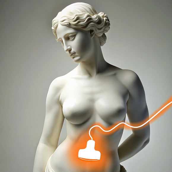
In women, the study helps to identify pathologies such as fibroids, cysts, endometriosis, inflammatory processes and tumor formations. In men, the procedure allows you to diagnose prostatitis, prostate adenoma and other diseases.
Benefits
Painlessness and safety
Ultrasound of the pelvic organs is a non—invasive and painless procedure.
High information content
The study provides detailed information about the condition of the internal organs of the pelvis.
Various visualization methods
The possibility of transabdominal, transvaginal and transrectal ultrasound allows you to choose the optimal method of examination.
Speed of implementation
The procedure takes a little time, and the results are available immediately after the study.
Preparation for ultrasound of the pelvic organs
An hour before the study, you should drink 1-1.5 liters of water to fill the bladder, which will improve the visualization of organs. For transvaginal ultrasound (in women) and transrectal ultrasound (in men), special training may be required, which the doctor will inform you about in advance.

Recommendations after ultrasound of the pelvic organs
After performing an ultrasound of the pelvic organs, the patient can immediately return to normal business. It is important to follow the doctor's recommendations, especially if pathologies have been identified that require observation or treatment.

Pelvic ultrasound procedure
For transabdominal ultrasound, the doctor applies a special gel to the skin of the abdomen and moves the sensor, assessing the condition of the pelvic organs through the abdominal wall. For transvaginal ultrasound in women, the sensor is inserted into the vagina, which allows you to get a more detailed image of the uterus and ovaries. For transrectal ultrasound in men, the sensor is inserted into the rectum, which makes it possible to examine the prostate gland in detail. The procedure usually takes about 20-30 minutes.

Врачи
Смотреть всех врачейIndications and contraindications
Indications
Pain and discomfort in the pelvic area
Pain in the lower abdomen, which may be associated with diseases of the internal organs of the pelvis.
Menstrual disorders in women
The study helps to identify the causes of cycle disorders such as fibroids, cysts and endometriosis.
Problems with urination in men
With difficulty urinating, pain and other symptoms indicating pathology of the prostate gland.
Pregnancy planning and monitoring
To assess the condition of the uterus and ovaries during pregnancy planning and during prenatal follow-up.
Expected effect
Accurate diagnosis
Ultrasound allows you to identify and evaluate diseases of the pelvic organs.
Monitoring of chronic diseases
Monitoring the condition of patients with chronic diseases of the pelvic organs.
Identification of hidden pathologies
Detection of diseases in the early stages, which allows you to start timely treatment.
Assessment of the state of reproductive organs
In women, the study helps in planning pregnancy and monitoring its course, in men — in assessing the condition of the prostate gland and seminal vesicles.
Similar referral activities
Ultrasound of the abdominal cavity
Ultrasound allows you to visualize internal organs such as the liver, gallbladder, pancreas, spleen, kidneys and other structures.
Ultrasound of the gallbladder
A study that allows you to assess the condition and functionality of the gallbladder, as well as identify pathologies such as stones, inflammatory processes and neoplasms.
Ultrasound of the mammary glands
A diagnostic method that allows you to evaluate the structure of breast tissue and identify pathological changes. Suitable for women of all ages and health conditions.
Ultrasound of the liver
Ultrasound examination of the liver allows you to visualize the structure and assess the condition of the liver.
Ultrasound of the kidneys and ureters
Ultrasound of the kidneys and ureters is a diagnostic study using ultrasound waves to visualize the kidneys and ureters, which allows you to identify stones, tumors, cysts, inflammatory and other pathological processes in these organs.
Ultrasound of the thyroid gland
A diagnostic method that allows you to evaluate the structure, size and blood supply of the thyroid gland, parathyroid glands and regional lymph nodes. It is indispensable in the diagnosis of diseases such as thyroiditis, thyrotoxicosis, nodular goiter and tumor processes.
How to reach


- Route from the «Sukharevskaya» metro station
- Route from the «Tsvetnoy Boulevard» metro station
From the metro station "Tsvetnoy Bulvar"
1 exit to the city, then left to the Garden Ring, at the crossing to the right, crossing the boulevard, one more crossing and at the traffic light to the left. The Olymp Clinic building is located overlooking the Garden Ring to the right of the crossing. Travel time is approximately 9 minutes. Landmark - sign Olymp Clini
From the metro station "Sukharevskaya"
Exit 3 from the metro and 640 meters straight ahead, the clinic will be on the right. Landmark - sign Olymp Clinic
Parking lot map
Exit 3 from the metro and 640 meters straight ahead, the clinic will be on the right. Landmark - sign Olymp Clinic