

Ultrasound of the mammary glands
A diagnostic method that allows you to evaluate the structure of breast tissue and identify pathological changes. Suitable for women of all ages and health conditions.
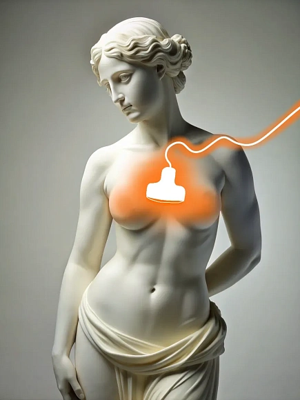
Ultrasound of the mammary glands allows you to examine in detail the tissue structure of the breast, revealing inflammation, cysts and tumors.
Special preparation for breast ultrasound is not required. It is optimal to undergo a study in the first days after the end of menstruation (5-9 days of the cycle), when the mammary glands are less susceptible to edema.
The procedure is usually performed in a sitting or lying position. The patient places her arm on the examined side behind her head to provide better access to the mammary gland. Gel is applied to the skin. The doctor runs the sensor over the entire surface of the breast, examining the glandular tissue, ducts and adjacent lymph nodes from different angles and in different projections. Images of tissues and vessels are recorded and stored digitally for further analysis.
A high-frequency ultrasound machine is used for ultrasound. Modern sensors have high resolution and allow you to get a detailed image of small structures.
After completion of breast ultrasound, the patient can immediately return to daily life. The results of the study are available immediately after the procedure and are transmitted to the attending physician for analysis.
Benefits
Safety
Ultrasound has no radiation effect and can be performed repeatedly.
High accuracy of soft tissue diagnostics
Ultrasound is especially effective for women with dense glandular tissue.
Absence of pain
The procedure does not cause discomfort and does not require special training.
A wide range of diagnosed conditions
The technique helps to diagnose a wide range of diseases.
Врачи
Смотреть всех врачейSimilar referral activities
Ultrasound of the abdominal cavity
Ultrasound allows you to visualize internal organs such as the liver, gallbladder, pancreas, spleen, kidneys and other structures.
Ultrasound of the gallbladder
A study that allows you to assess the condition and functionality of the gallbladder, as well as identify pathologies such as stones, inflammatory processes and neoplasms.
Ultrasound of the pelvic organs
Ultrasound allows you to visualize and assess the condition of the internal organs of the pelvis in women and men. In women, the study includes examination of the uterus, ovaries and fallopian tubes, in men — the prostate gland and seminal vesicles.
Ultrasound of the liver
Ultrasound examination of the liver allows you to visualize the structure and assess the condition of the liver.
Ultrasound of the kidneys and ureters
Ultrasound of the kidneys and ureters is a diagnostic study using ultrasound waves to visualize the kidneys and ureters, which allows you to identify stones, tumors, cysts, inflammatory and other pathological processes in these organs.
Ultrasound of the thyroid gland
A diagnostic method that allows you to evaluate the structure, size and blood supply of the thyroid gland, parathyroid glands and regional lymph nodes. It is indispensable in the diagnosis of diseases such as thyroiditis, thyrotoxicosis, nodular goiter and tumor processes.
How to reach


- Route from the «Sukharevskaya» metro station
- Route from the «Tsvetnoy Boulevard» metro station
From the metro station "Tsvetnoy Bulvar"
1 exit to the city, then left to the Garden Ring, at the crossing to the right, crossing the boulevard, one more crossing and at the traffic light to the left. The Olymp Clinic building is located overlooking the Garden Ring to the right of the crossing. Travel time is approximately 9 minutes. Landmark - sign Olymp Clini
From the metro station "Sukharevskaya"
Exit 3 from the metro and 640 meters straight ahead, the clinic will be on the right. Landmark - sign Olymp Clinic
Parking lot map
Exit 3 from the metro and 640 meters straight ahead, the clinic will be on the right. Landmark - sign Olymp Clinic